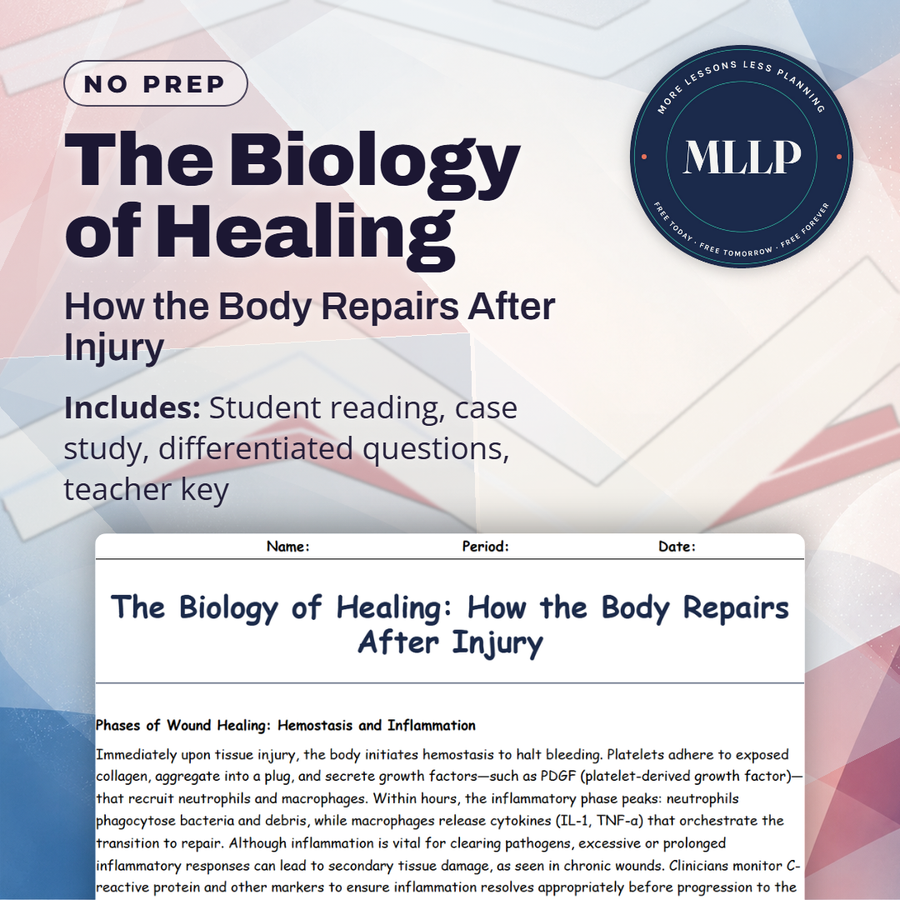
The Biology of Healing: How the Body Repairs After Injury
Quality reviewed September 2026
Facts verified September 9, 2026
- Subject
- Science
- Grades
- Grades 9-12
- Length
- 7-page student worksheet
- Time
- About one 50-minute class period
- Great for
- Sub plans and independent work
- Includes
- Reading passage, comprehension questions + full answer key
- Format
- Print-and-go PDF
What students do
- Analyze the three phases of wound healing from hemostasis to remodeling
- Explain how angiogenesis rebuilds blood supply in injured tissue
- Evaluate clinical interventions including hyperbaric oxygen and dermal substitutes
- Trace Corporal Lee's healing from field dressing to return to duty
What it covers
- Hemostasis, inflammation, and immune cell recruitment
- Angiogenesis and granulation tissue formation
- Scar formation and long-term remodeling
Start reading this worksheet
Phases of Wound Healing: Hemostasis and Inflammation
Immediately upon tissue injury, the body initiates hemostasis to halt bleeding. Platelets adhere to exposed collagen, aggregate into a plug, and secrete growth factors, such as PDGF (platelet-derived growth factor), that recruit neutrophils and macrophages. Within hours, the inflammatory phase peaks: neutrophils phagocytose bacteria and debris, while macrophages release cytokines (IL-1, TNF-α) that orchestrate the transition to repair. Although inflammation is vital for clearing pathogens, excessive or prolonged inflammatory responses can lead to secondary tissue damage, as seen in chronic wounds. Clinicians monitor C-reactive protein and other markers to ensure inflammation resolves appropriately before progression to the next healing stage.
Proliferation and Angiogenesis: Building New Tissue
Around day 3 post-injury, the body shifts from defense to construction. Angiogenesis, stimulated by VEGF (vascular endothelial growth factor), sprouts new capillaries into the wound bed, forming granulation tissue rich in fibroblasts and extracellular matrix. Fibroblasts deposit type III collagen, creating a scaffold for cellular migration. Epithelialization follows, as keratinocytes migrate across the wound surface to reestablish the skin barrier. In organs like the liver, parenchymal cells can regenerate original architecture; in skin and muscle, however, repair leads to scar tissue, which restores continuity but lacks full biomechanical function. Proper oxygenation, nutrition, and absence of infection are critical for robust proliferation.
Remodeling and Scar Formation
From week 3 onward, the wound enters the remodeling phase, which can last months to years. Matrix metalloproteinases (MMPs) degrade excess extracellular matrix, while fibroblasts transition to myofibroblasts, contracting the wound and aligning collagen fibers along tension lines to increase tensile strength. Type III collagen is replaced by stronger type I collagen, and new blood vessels regress through apoptosis. Although remodeled tissue recovers up to 80 percent of the original strength, it remains less elastic and more prone to reinjury. Interventions (such as silicone gel sheeting, pressure garments, and corticosteroid injections) help modulate excessive scar formation (keloids) and optimize functional outcomes.
Like what you read? The full reading, the case study, and every question set are in the free download below, along with the answer key.
Why this matters
Hemostatic gauze packed into Corporal Lee's fresh wound stops the bleeding in about ten minutes, and four days later a Doppler ultrasound at the surgical unit confirms new blood vessels already growing into the injured tissue. That timeline, not a diagram of cell types, is what makes healing concrete for students.
What changes once students work through the full case, from hemostasis through inflammation and granulation tissue to a dermal substitute and eight weeks of remodeling, is that healing stops looking automatic. They start asking why the order of those phases matters at all.
From one teacher to another
Corporal Lee's calf laceration carries this worksheet because the timeline is concrete. A field medic packs the wound with hemostatic gauze and reaches hemostasis in about ten minutes, then on day four a Doppler ultrasound at the surgical unit confirms new vessels growing into the tissue. From there the reading walks through inflammation, granulation tissue, a bioengineered dermal substitute, and eight weeks of remodeling. My students like that cells and surgeons show up in the same story. The questions ask them to place each intervention inside the three healing phases and explain why the order matters.
What you get
Download
Where should I send the next one?
Everything here is free and it stays that way. Leave your email and I'll send you the new lessons as they go up. You can unsubscribe from any one of them.
Related resources

Memorial Day and Environmental Impact: Waste, Travel, and Outdoor Recreation

Memorial Day: Honoring Sacrifice and Remembering History

Memorial Landscapes: How Weather, Erosion, and Time Shape War Memorials
The Forensics of War: Identifying the Fallen

The Psychology of Grief, Memory, and Memorializing
The Science of Combat Injuries: How the Body Responds to Trauma
Every lesson on this site is free to download and use. If one of them saved you a prep period, you can chip in a few bucks toward my three kids' daycare fund. That's genuinely where tips go.