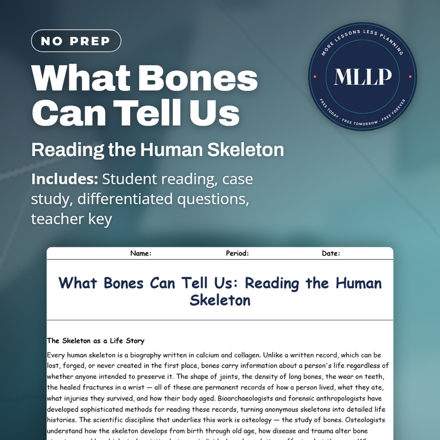
What Bones Can Tell Us: Reading the Human Skeleton
Quality reviewed September 2026
Facts verified September 21, 2026
- Subject
- Social Studies
- Grades
- Grades 9-12
- Length
- 8-page student worksheet
- Time
- About one 50-minute class period
- Great for
- Sub plans and independent work
- Includes
- Reading passage, comprehension questions + full answer key
- Format
- Print-and-go PDF
What students do
- Sort the skeletal markers, from enthesopathies to Harris lines, by what kind of life history each one reveals
- A skeleton's pelvis and skull carry the most reliable clues to biological sex that osteologists ever get
- Explain why the York skeleton's dental work, malnutrition markers, and labor injuries do not fit one simple life story
- Compare a healed fracture to an unhealed one and what each suggests about survival
What it covers
- How osteologists estimate age and biological sex from growth plates, joint wear, and pelvic shape
- What enthesopathies, Harris lines, and porotic hyperostosis reveal about labor and childhood nutrition
- Why a healed fracture and an unhealed one tell different stories about survival and cause of death
Start reading this worksheet
The Skeleton as a Life Story
Every human skeleton is a biography written in calcium and collagen. Unlike a written record, which can be lost, forged, or never created in the first place, bones carry information about a person's life regardless of whether anyone intended to preserve it. The shape of joints, the density of long bones, the wear on teeth, the healed fractures in a wrist: all of these are permanent records of how a person lived, what they ate, what injuries they survived, and how their body aged. Bioarchaeologists and forensic anthropologists have developed sophisticated methods for reading these records, turning anonymous skeletons into detailed life histories. The scientific discipline that underlies this work is osteology, the study of bones. Osteologists understand how the skeleton develops from birth through old age, how disease and trauma alter bone structure, and how biological variation between individuals and populations affects what they see. When a bioarchaeologist examines a skeleton, they are not guessing. They are applying well-established biological principles to physical evidence, producing estimates and conclusions that can be tested, refined, and compared to findings from other sites.
Age, Sex, and Stature: The Biological Profile
The first task when examining an unknown skeleton is constructing a biological profile, a set of basic descriptors that narrow down who this person was. Estimating age at death is one of the most reliable assessments osteologists make. In children and adolescents, the timing of tooth eruption and the fusion of growth plates at the ends of long bones follow predictable developmental schedules. A skeleton with all its growth plates still open and primary teeth present almost certainly belonged to a young child. By contrast, in adults, age estimation becomes less precise and relies on degenerative changes: wear on joint surfaces, changes in the density of the pubic symphysis (where the two halves of the pelvis meet), and the degree of fusion of cranial sutures. Estimating biological sex from skeletal remains is most reliable from the pelvis and skull. The female pelvis evolved to accommodate childbirth and has a wider, more open shape than the male pelvis. The male skull tends to have more pronounced brow ridges, a larger mastoid process behind the ear, and a more angular jaw. These differences are reliable in adults but less so in children, whose skeletons have not yet developed sexually dimorphic features. Stature can be estimated from the length of long bones (particularly the femur and tibia) using population-specific regression formulas that account for the relationship between limb length and overall height.
Markers of Hard Work and Hard Times
Beyond the basic biological profile, bones record the texture of a life. Enthesopathies are bony projections or roughened areas that develop where muscles and tendons attach to bone. They grow more pronounced with heavy, repetitive use. A medieval laborer who spent decades carrying loads would develop dramatically different enthesopathies than a scribe who spent the same years sitting at a desk. By mapping these markers across a skeleton, bioarchaeologists can make educated inferences about the physical demands of an individual's occupation. Nutrition leaves its mark on bones as well. Episodes of malnutrition during childhood cause horizontal lines to form in the dense cortex of long bones, visible on X-rays as Harris lines. Severe vitamin D deficiency produces rickets, a condition that bows the leg bones and deforms the pelvis. Scurvy, caused by vitamin C deficiency, leaves characteristic lesions on the surface of bones where blood vessels ruptured. Anemia can cause the skull's outer table to become porous and pitted, a condition called porotic hyperostosis. Together, these markers allow researchers to reconstruct not just an individual's diet, but the food security of entire ancient communities.
Like what you read? The full reading, the case study, and every question set are in the free download below, along with the answer key.
Why this matters
A lot of students assume written records are the most reliable way to learn about someone's life, but this reading argues that bone can outlast and outtell paper, since a skeleton records things no one intended to write down. Dr. Vasquez's case study of skeleton YK-247 shows why reading multiple markers together matters: dental work suggests money, malnutrition markers suggest early poverty, and labor injuries suggest a lifetime of physical work, and no single marker would have told the full story alone. Students who are drawn to medicine, anthropology, or forensic science get to see how physical evidence, read carefully, can reconstruct a life story more completely than most surviving documents ever could.
From one teacher to another
Bones outlast paper records, and this reading treats a skeleton as a biography written in calcium and collagen. It walks through how osteologists build a biological profile, estimating age from growth plates and tooth eruption in the young and joint wear in adults, and estimating biological sex mainly from the shape of the pelvis and skull. From there it covers enthesopathies, the bony ridges that build up at muscle attachment points from decades of repetitive labor, along with Harris lines and porotic hyperostosis, which record childhood malnutrition and anemia. The final passage explains how healed and unhealed fractures tell different stories about survival and cause of death. The case study follows Dr. Elena Vasquez as she reads a York skeleton's teeth, spine, and shoulders for a life story that does not fit into one simple category.
What you get
Download
Where should I send the next one?
Everything here is free and it stays that way. Leave your email and I'll send you the new lessons as they go up. You can unsubscribe from any one of them.
Related resources
Battlefield Archaeology: Uncovering the Cost of War
Bones, DNA & Forensic Archaeology: An Overview
Facial Reconstruction: Putting a Face on the Past
Forensic Archaeology: Solving Modern Crimes with Ancient Methods
_COVER.png)
Isotope Analysis: You Are What You Eat (and Where You Lived)
Mummies: Preserved Bodies and What They Teach Us
Every lesson on this site is free to download and use. If one of them saved you a prep period, you can chip in a few bucks toward my three kids' daycare fund. That's genuinely where tips go.